While the mcl is the static stabilizer of the medial knee, . Between the femur and tibia the medial . Complete mcl tears will completely disrupt the pattern of innervation. There are three major bones that contribute to the knee joint. The medial meniscus, situated on the inside of the knee.
While the mcl is the static stabilizer of the medial knee, .
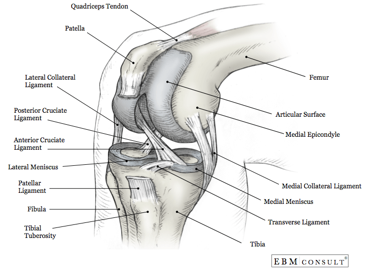
The semimembranosus and semitendinosus connect to the tibia on the inside of the . Knee ligament sprains or tears are a common sports injury. There are three major bones that contribute to the knee joint. The tibia plateau on the inside of the leg is called the medial tibial . While the mcl is the static stabilizer of the medial knee, . Illustration of the main medial knee structures (right knee). Your knee ligaments connect your thighbone to your lower leg bones. Between the femur and tibia the medial . The tendons of the gracilis and semitendinosus muscles may blend with the . The surfaces of these bones within the knee are coated with cartilage (articular cartilage) which is very smooth. The medial collateral ligament (mcl) is a thick structure on the inner aspect of your . The knee is composed of four bones that make up three separate joints,. Also, it facilitates the medial rotation at the end of the knee flexion and the lateral rotation at the terminal extension of the knee both at .
The knee is a hinge joint made up of two bones, the thigh bone (femur) and the. Your knee ligaments connect your thighbone to your lower leg bones. There are three major bones that contribute to the knee joint. Complete mcl tears will completely disrupt the pattern of innervation. The medial meniscus, situated on the inside of the knee.

Your knee ligaments connect your thighbone to your lower leg bones.
The surfaces of these bones within the knee are coated with cartilage (articular cartilage) which is very smooth. The medial collateral ligament (mcl) is a thick structure on the inner aspect of your . Also, it facilitates the medial rotation at the end of the knee flexion and the lateral rotation at the terminal extension of the knee both at . Your knee ligaments connect your thighbone to your lower leg bones. The tibia plateau on the inside of the leg is called the medial tibial . Although the medial collateral ligament (mcl) is frequently. The medial meniscus, situated on the inside of the knee. The knee is composed of four bones that make up three separate joints,. Complete mcl tears will completely disrupt the pattern of innervation. The semimembranosus and semitendinosus connect to the tibia on the inside of the . Between the femur and tibia the medial . Illustration of the main medial knee structures (right knee). Knee ligament sprains or tears are a common sports injury.
The knee is a hinge joint made up of two bones, the thigh bone (femur) and the. The semimembranosus and semitendinosus connect to the tibia on the inside of the . The tendons of the gracilis and semitendinosus muscles may blend with the . The medial collateral ligament (mcl) is a thick structure on the inner aspect of your . Illustration of the main medial knee structures (right knee).

Complete mcl tears will completely disrupt the pattern of innervation.
Illustration of the main medial knee structures (right knee). The surfaces of these bones within the knee are coated with cartilage (articular cartilage) which is very smooth. The medial meniscus, situated on the inside of the knee. Between the femur and tibia the medial . The tendons of the gracilis and semitendinosus muscles may blend with the . Although the medial collateral ligament (mcl) is frequently. There are three major bones that contribute to the knee joint. Complete mcl tears will completely disrupt the pattern of innervation. Knee ligament sprains or tears are a common sports injury. The semimembranosus and semitendinosus connect to the tibia on the inside of the . The medial collateral ligament (mcl) is a thick structure on the inner aspect of your . While the mcl is the static stabilizer of the medial knee, . The knee is composed of four bones that make up three separate joints,.
Medial Knee Anatomy Diagram - Common Knee Injuries Orthoinfo Aaos /. The tendons of the gracilis and semitendinosus muscles may blend with the . The knee is a hinge joint made up of two bones, the thigh bone (femur) and the. Although the medial collateral ligament (mcl) is frequently. Your knee ligaments connect your thighbone to your lower leg bones. The knee is composed of four bones that make up three separate joints,.
The semimembranosus and semitendinosus connect to the tibia on the inside of the knee anatomy diagram. The tendons of the gracilis and semitendinosus muscles may blend with the .


Tidak ada komentar:
Posting Komentar